preprint update: Fountains are the first structures to fold the genome in developing zebrafish embryos

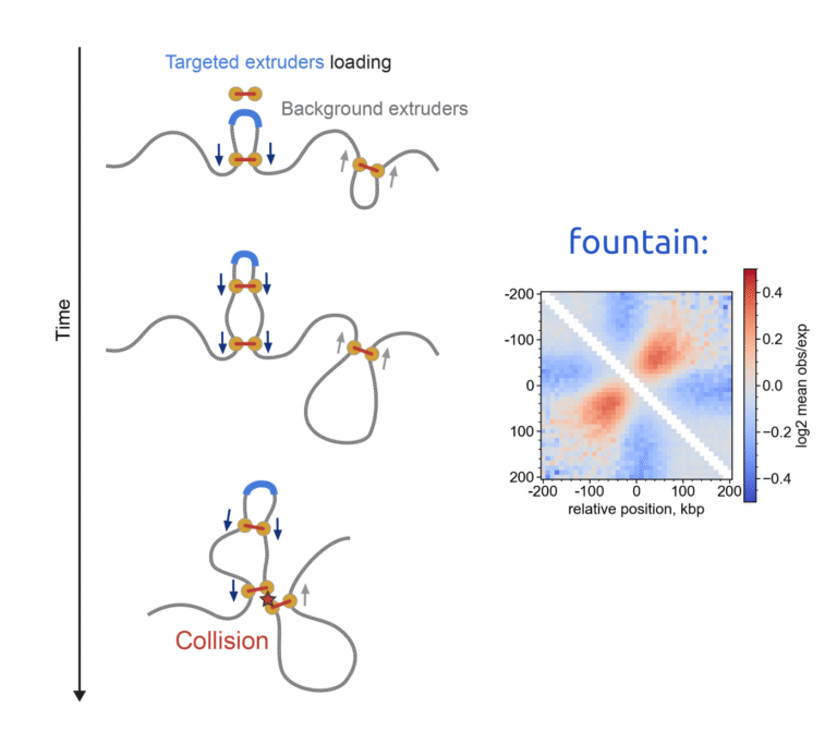
In this update, I show that chromatin “fountains” form at enhancers not only in early zebrafish embryos but also during mitotic exit in mouse embryonic stem cells (mESCs). This is exciting because fountains appear to be a universal early feature of genome folding and likely reflect cohesin loading at enhancers.

My summary below focuses on the polymer-simulation side. To reproduce the characteristic fountain signature in Hi-C/Micro-C, you need (i) targeted loading of loop extruders (e.g., cohesin) at specific genomic sites—likely enhancers—and (ii) background obstacles that impede cohesin’s reeling. Together, these ingredients generate the fountain-like enrichment pattern in Hi-C.

The original preprint appeared on bioRxiv in 2023, and we’re now sharing a revised version. More details:

https://www.biorxiv.org/content/10.1101/2023.07.15.549120v2.full


Share the Post:

Related Posts

The story that features in NAR

A very curious part of my PhD work on Dictyostelium discoideum chromatin has been published in Nucleic Acid Research this week. My part was designing a 1D extrusion and polymer model to explain why the “bright dots” are happening at the convergent gene positions (CGPs). 

Read More

preprint: Deciphering the 3D genome organization across species from Hi-C data

I’m pleased to share the results of a project I started during my graduate studies, now beautifully completed by my colleagues.

The study applies a sophisticated method to construct joint embeddings of DNA sequences and Hi-C chromatin organization maps within a shared space. This framework allows for the exploration of chromatin structures with distinct functional characteristics, such as insulation domains, loops, and fountains. Intriguingly, it also enables us to identify DNA sequences associated with specific structural features and even modify chromatin maps by ‘adding’ these elements.

One particularly striking result is the construction of a ‘tree of life’ for chromatin spatial organization. While it bears some similarity to traditional phylogenetic trees based on DNA sequences, there are notable differences that reflect significant evolutionary shifts in mechanisms of chromatin folding.

https://www.biorxiv.org/content/10.1101/2024.11.14.623548v2

Read More